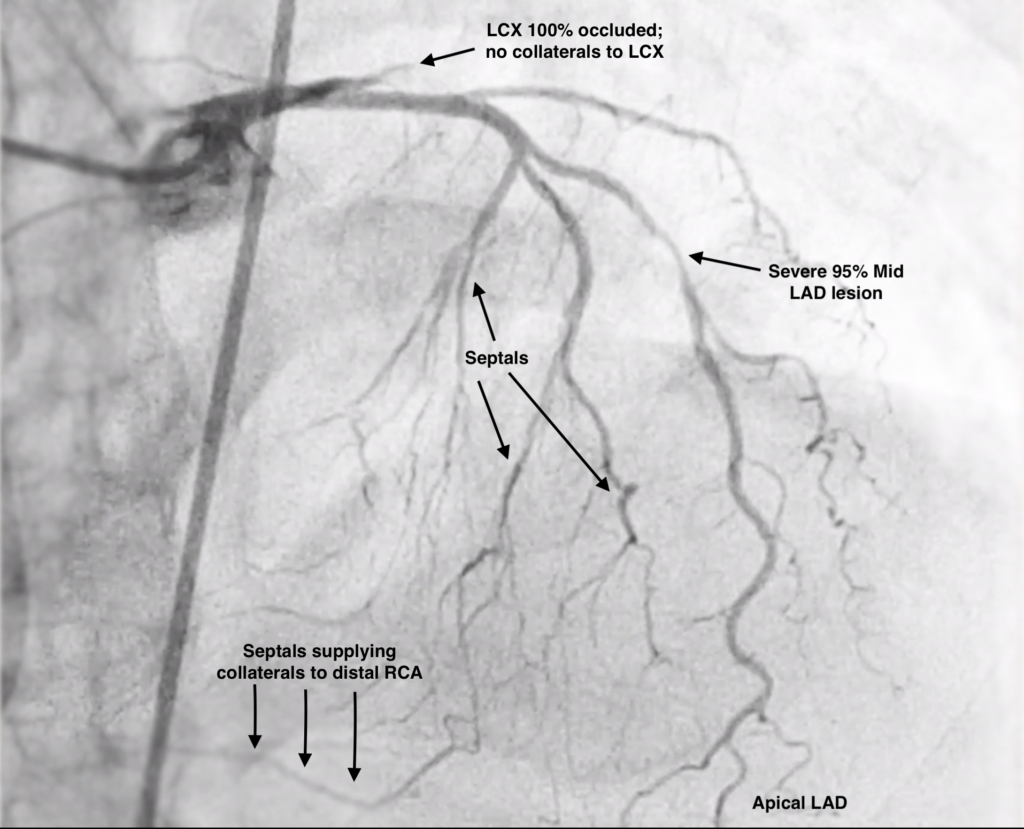
The correct answers here are: Severe disease for LAD, Total Occlusion for LCX, Total Occlusion for RCA, and RCA is Filled by Collaterals. The Left Main appears Normal in this view
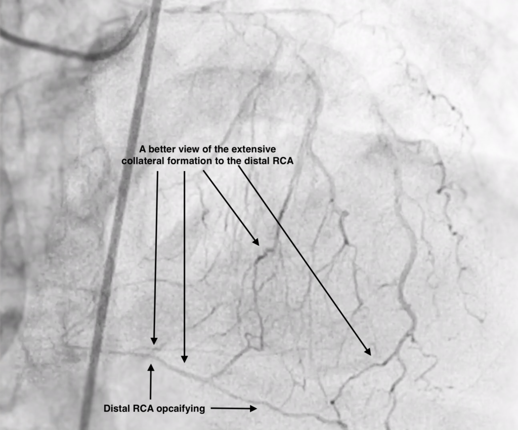
The first step in interpreting this angiogram is to orient yourself. This is a RAO Cranial projection. There are large septals coming across the screen indicating the LAD is the main vessel we see. Sometimes it can be challenging to even accurately identify the LAD, especially when the septal branches are larger in size. In this case, there is a large septal almost masquerading as an LAD, but the only true vessel that goes to the apex is to the right so that is the LAD. A key finding is to observe that the LAD septal branches are filling the right coronary artery distally so the RCA has a total occlusion.
Finally, the LCX stumps off and there is contrast staining where the LCX pathway would normally be. There also do not appear to be collaterals going to the LCX suggesting this is an acute lesion and likely a lateral STEMI
In situations such as this, it can be challenging to identify what the culprit lesion is. It is important to review the clinical information you have at hand, such as the patient’s electrocardiogram and echocardiogram if you have one. Collateral formation can help you tease apart acute vs chronic lesions such as in this angiogram.